
1-15 Lexington Rd, Underwood, QLD, AU, 4119
SEARCH

What can an ultrasound of a feline gallbladder tell me?

Biochemistry and clinical signs may strongly suggest hepatobiliary involvement, but they rarely localise the problem or explain its mechanism. Ultrasound of the feline gallbladder allows its direct assessment, including the bile ducts and surrounding structures, to distinguish primary biliary disease from secondary change linked to hepatic, pancreatic or intestinal pathology.
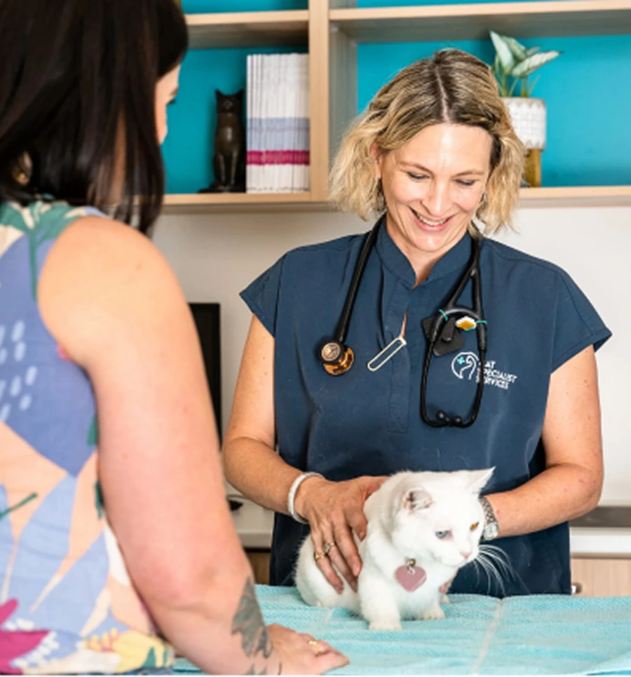
At Cat Specialist Services, we work exclusively with feline patients, and we’re committed to supporting general practice vets and nurses through complex cases. When it comes to the gallbladder, findings shouldn’t ever be read in isolation. Sludge, wall changes or ductal dilation can have very different clinical significance depending on the wider presentation.
Establish the baseline first
Familiarity with the normal feline gallbladder is essential before subtle or early change can be recognised. Without a clear baseline, normal anatomical variation can be mistaken for pathology, or early disease can be overlooked. In cats, the gallbladder is typically located between the quadrate and right medial liver lobes and appears narrow and elongated, with a thin, smooth wall and an anechoic lumen when bile is normal.
Consistency in measurement technique is critical when assessing the biliary tree, particularly the common bile duct. Measurements taken at the porta hepatis will provide the most reliable reference point, as this region is less affected by transient changes in duodenal filling or patient positioning. Using the same anatomical landmarks across serial scans allows subtle progression or resolution of ductal changes to be identified with greater confidence.
Gallbladder imaging is often affected by gas, rib shadowing or poor acoustic windows, particularly in cats with gastrointestinal disease. Small adjustments in positioning and scanning planes can improve visualisation and reduce the risk of artefact being misinterpreted as true intraluminal or wall pathology.
Interpreting gallbladder contents on ultrasound
Clinically, not all echogenic material within the gallbladder is abnormal. Fine sludge often layers dependently and shifts position when the patient is moved, reflecting thickened bile rather than structural disease. In contrast, organised mucus forms more structured patterns that remain fixed within the lumen and may create stellate or striated appearances. These non-mobile patterns suggest abnormal bile composition and early mucocele type change, even without complete obstruction.
Features originating from the gallbladder wall rather than its contents suggest a different disease process. For example, wall thickening, layering or a hypoechoic halo is more consistent with inflammation or compromised perfusion than simple bile stasis. When these findings are present, particularly alongside reduced appetite, vomiting, abdominal discomfort or systemic illness, they are more likely linked to inflammatory biliary disease than incidental sludge.
Occasionally, highly echogenic foci with acoustic shadowing may be identified within the gallbladder lumen or wall. Although these findings can indicate mineralisation or cholelith formation, they are uncommon in cats and are not always clinically significant. Without supporting evidence such as ductal obstruction, pain or biochemical disturbance, isolated shadowing structures should be interpreted cautiously to avoid unnecessary escalation.

Signs of impaired bile flow and identifying the source
The most reliable ultrasound clue indicating impaired bile flow is seen in the common bile duct. Dilatation beyond expected limits, particularly when accompanied by upstream ductal distension and a downstream return to normal width, supports obstruction distal to the liver rather than primary hepatic dysfunction. This finding provides greater diagnostic value than gallbladder contents alone.
Even a feline gallbladder that appears normal doesn’t necessarily rule out significant biliary disease. Following the bile duct from the porta hepatis to its distal insertion into the duodenal at the level of the duodenal papilla is very important as this can identify wall irregularities, luminal debris or focal narrowing that may otherwise be missed.
Careful assessment of the papillary region is also important, as disease here can obstruct bile flow without obvious gallbladder changes.
However, not all biliary obstructions originate within the biliary system itself. Pancreatic inflammation, mass lesions or adjacent neoplastic processes can externally compress or distort the bile duct, causing a secondary obstruction pattern.
Using ultrasound findings to inform case management
Ideally, gallbladder ultrasound findings should guide clinical decisions around management, further investigation and urgency. In practice, this includes:
- Context matters with sludge
Gallbladder sludge is a common ultrasound finding and, on its own, does not always indicate infection or justify antimicrobial therapy without supporting clinical or biochemical evidence. - Using ultrasound to inform further investigation
Imaging helps determine whether diagnostics such as bile sampling or liver aspirates are likely to change management, rather than adding unnecessary intervention. - Urgency depends on the whole picture
The clinical significance of ultrasound findings increases when paired with systemic illness or changes such as ductal obstruction, gallbladder wall thickening or peritoneal fluid.
The bigger biliary picture
Essentially, a feline gallbladder ultrasound can reveal a great deal for veterinarians, from highlighting obstruction risk to characterising intraluminal material and guiding next steps. Interpretation, however, will always rely on context.
At Cat Specialist Services, our internal medicine team works closely with referring vets to support complex biliary cases, whether that involves image review, outpatient ultrasound, recommendations for further investigation or ongoing case management. Referrals and discussions are always welcome.
Refer here.
Are you worried about your cat’s health?
If you are concerned about your cat, please reach out to our friendly team. Call us on 1300 228 377 or fill out the contact form below. In an emergency, please contact your local vet.
For vets – to refer a patient or book a telehealth consultation, please call us on 1300 228 377, book through the portal or fill in the form.
Meet The Author
Dr Rachel Korman
Specialist in Feline Internal Medicine
BVSc MANZCVS (Internal Medicine) FANZCVS (Feline Medicine)
Dr. Korman’s childhood love for cats led her to veterinary medicine, and early on, it was clear that feline care was her calling. After graduating from the University of Queensland in 2000, she worked in small animal and feline-only practices across Australia and the UK.
She received a Senior Clinical Training Scholarship from the Feline Advisory Bureau (now International Cat Care) at the University of Bristol, where she researched feline infectious and haematological diseases, and in 2018, became a Fellow of the Feline Medicine Chapter of the Australian New Zealand College of Veterinary Scientists and a registered Feline Medicine specialist.
Frequently Asked Questions
What can an ultrasound of a feline gallbladder tell me?
It allows direct assessment of the gallbladder, bile ducts and surrounding structures to help distinguish primary biliary disease from secondary changes linked with liver, pancreas or intestinal pathology.
How is the gallbladder normally visualised on ultrasound?
A normal feline gallbladder appears narrow and elongated with a thin smooth wall and anechoic (clear) lumen when bile is uncomplicated—this baseline helps recognise subtle abnormalities.
What does it mean if there’s echogenic material, like sludge, seen in the gallbladder?
Echogenic material can represent thickened bile (sludge) and may not necessarily indicate disease on its own; interpretation depends on the wider clinical context including clinical signs and bloodwork.
Can ultrasound detect bile duct obstruction or impaired bile flow?
Yes — ultrasound can show dilation of the common bile duct and other changes that point to impaired bile flow or obstruction, which provides valuable diagnostic clues beyond gallbladder appearance alone.
How are ultrasound findings used to guide further management?
Results help veterinarians decide whether additional diagnostics (like bile sampling) are needed, assess urgency, and determine next steps in case management by correlating imaging with clinical and biochemical data.
Patient Stories
Our commitment to feline health is best seen in the stories of the cats we’ve had the privilege of treating. These journeys highlight the dedication, expertise and compassion that drive us.
Each patient faced unique challenges, and it was our honour to offer high-quality care and help them regain strength and happiness. We value the deep bonds with cats and their owners, knowing the difference we make in their lives.
Kitty, a lymphoma survivor
We first met Kitty after she was referred to CSS for the evaluation of a large intestinal mass
Poppi finds her feet again
Following surgery to remove a hairball obstruction, Poppi was referred to us at CSS as she had not bounced back as expected
Rusty and his ongoing chronic rhinitis journey
Lifelong nasal issues, including persistent discharge and breathing noise, have been a bit of a thorn in Rusty’s side.
Lando’s long game with cryptococcus
Lando was referred to CSS because he had a swelling under his jaw that wouldn’t go away.
Popeye coughs up the culprit
Ares is a handsome young Maine Coon who was referred to CSS after presenting at AES with sudden lameness in his back leg.
Key Takeaways

Gallbladder ultrasound enables veterinarians to evaluate the gallbladder, bile ducts, and nearby organs to better understand hepatobiliary disease in cats.
Features like sludge, wall thickening, or duct dilation can have different clinical meanings depending on symptoms, blood tests, and overall health findings.
Ultrasound helps determine whether further tests such as bile sampling or additional imaging are necessary and whether a condition requires urgent intervention.
Cat Specialist Services Reviews  on
on 
Tanya Bell
6 months ago
We transported our cat here from another animal hospital, as his condition was too complex for them. Our pet was given the highest quality of care, multiple investigations/procedures being able to be done at the one place a godsend. We were kept up to date frequently throughout his stay. All questions & options explained thoroughly. Being able to have private visits certainly helped us and our cat too. I would highly recommend Cat Specialist Services at Underwood. Amazing staff from reception, vet nurses, vets & specialists. Oh, and yes he is recovering wonderfully at home.
Melinda Cox
A month ago
So very happy we were asked to come here for advice for our NORBIT. He has had 2 x UTI blockages within weeks of each other since turning 6. Surgery seemed to be his only option and he didn’t feel it was very fair on him if we didn’t at least exhaust all other avenues first.
Dr Allison was very thorough in her tests and has let us know she would like to treat the cause of the problem. Great news.
It may lead to removal of crystals in his bladder if his diet doesn’t let him pass them naturally.
At least this surgery won’t be as severe at taking his penis off him – so as his fur parents – we are very happy with this.
We await further tests to see what’s in store for him.
NORBIT will now be a patient of the clinic as we are so far very happy with his treatment thus far.
Lesley Rosekrans
A month ago
Hi has only been a week since losing my beautiful Peaches but can’t thank Dr Cindy, Maree and Jackie enough for the tender care they gave her and they after care they gave me. I sincerely thank them and can’t recommend the Clinic highly enough they are such caring people. Peaches I’m happier times.
Madison McEwan
A month ago
I cannot thank the entire CSS team enough and especially Dr Cindy for the care they provided for our boy with a urinary blockage, and we will be taking both our cats here from now on. The clinic is exceptionally clean and calming compared to all other clinics we’ve been too, and all staff from the reception to the nurses/vets were so supportive during a stressful time. I knew from the first minute that I got to see him after he was transferred that he was in the best place, and that the staff genuinely care about their well-being and positive long term outcomes. We were given regular updates and full transparency, so much information including QR video codes on how to administer medications, along with print outs and emails about how to best help avoid future issues or recurrence. Our boy got to smooch on everyone and was beyond well looked after. Thank you again, you all made an incredibly stressful situation actually manageable and calming.
Astrid El Gamal
6 months ago
Dr Wan-Ju has been treating my baby since her first seizure in November 2025 and she has been fantastic.
Everybody at the clinic is so friendly and caring and I know my baby is in good hands.
Thank you team!
Deeba M
3 months ago
Dr Alison Jukes from CSS Underwood is an AMAZING feline physician. She is kind, professional, thorough and our cat is blessed to have her on his side. She even was able to save us money and invasiveness by being skilled to the level of doing ultrasound on two separate occasions without our cat needing to go under full sedation. Our other cat was recently seen by Dr Jukes as well, and she made sure our cat’s heart was ok. I also want to thank the lovely Jacqueline and Mairead from customer care. They are very kind each time I see them and call up. Sending thanks from our family, including Smokey and Floozy!
Judith Dionysius
A month ago
My whole experience with CSS was positive – from the first email I received giving lots of information regarding the process of radioactive iodine treatment to the expected costs, to checking my cat in for the treatment, boarding for 2 weeks after the treatment and picking her up to come home. They didn’t push for any tests that were not necessary.
Janelle Wieden
5 months ago
We never knew this service existed but we are very grateful we were referred to them. Our vet Cindy was really great, she made us feel at ease during a very stressful time. We got the sense straight away the Leo was in good hands. This service made us feel better knowing that they specialise is cats only. Would highly recommend their services
Georgia Carter
5 months ago
My Lani finished her I-131 treatment today and I am beyond impressed with the way she was looked after. I am by definition a neurotic pet owner and sending my fur child away for a week was seriously daunting.
I was given daily updates about how kitty was eating, toileting and behaving. This really put my mind at ease. Vets and vet nurses were so accomodating and allowed Lani to have her favourite foods from home prepared her favourite way.
Her vet was so thorough when explaining her treatment and condition to me.
The ladies at reception were so friendly and understanding.
I honestly cannot recommend this clinic enough and I am so thankful for the positive experience Lani and I have had.
Lauren Woodward
A year ago
My sweet ginger boy Louis was hospitalised at CSS for a Urinary Obstruction. Dr Cindy and all the reception staff were amazing and empathetic. I would often ring up with questions post procedure and Dr Cindy would make time to talk and explain things to me
Service Locations
Cat Specialists accepts referrals for cats across Brisbane and surrounding regions, working closely with local veterinary clinics to provide specialist diagnosis, treatment, and ongoing management.


Contacting Us
Pet Owners
If you would like to talk about treatment for your cat, call us on 1300 228 377 or fill in the form.
In an emergency, please contact your local vet.
Vets and Nurses
To refer a patient or book a telehealth consultation please call us on 1300 228 377, book through the portal or fill in the form. For advice calls, please see the guidelines here.


 Monday-Friday: 8 am-6 pm
Saturday/Sunday - Closed